A hyperintensity or T2 hyperintensity is an area of high intensity on types of magnetic resonance imaging (MRI) scans of the brain of a human or of another...
10 KB (1,200 words) - 16:25, 19 March 2025

Leukoaraiosis (redirect from White matter hyperintensity)
young adults. On MRI, leukoaraiosis changes appear as white matter hyperintensities (WMHs) in T2 FLAIR images. On CT scans, leukoaraiosis appears as hypodense...
10 KB (989 words) - 00:39, 31 August 2023

structure and function. In particular, methamphetamine appears to cause hyperintensity and hypertrophy of white matter, marked shrinkage of hippocampi, and...
170 KB (16,612 words) - 21:29, 29 May 2025

symptom onset to develop. Imaging by MRI typically demonstrates areas of hyperintensity on T2-weighted images. To minimise the risk of this condition developing...
29 KB (3,099 words) - 15:34, 26 May 2025
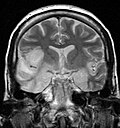
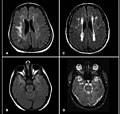
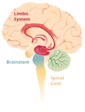
memory loss, seizures, and behavioral anomalies. MRI imaging reveals T2 hyperintensity in the structures of the medial temporal lobes, and in some cases, other...
29 KB (2,539 words) - 18:13, 28 January 2025

MRI (b) of left wrist showing necrotizing fasciitis. There is diffuse hyperintensity with irregular enhancement of the deep fascia (asterisks). The arrows...
50 KB (4,988 words) - 18:04, 23 May 2025
ARIA-E Edema FLAIR hyperintensity confined to sulcus and/or cortex/subcortical white matter in one location < 5 cm FLAIR hyperintensity 5 to 10 cm, or more...
10 KB (779 words) - 15:29, 15 April 2025

thickened, darker signal of the junctional zone, one will often see foci of hyperintensity (bright spots) on the T2 weighted scans representing small cystically...
41 KB (4,758 words) - 16:05, 13 May 2025

people with bipolar disorder have higher rates of deep white matter hyperintensities. Functional MRI findings suggest that the ventricular prefrontal cortex...
164 KB (18,063 words) - 23:31, 26 May 2025
hypointense area is thought to be caused by the excess iron while the central hyperintensity is possibly a result of gliosis. Guillerman, R. Paul (2000). "The Eye-of-the-Tiger...
2 KB (169 words) - 21:06, 25 May 2025

been called "cortical ribboning" or "cortical ribbon sign" due to hyperintensities resembling ribbons appearing in the cortex on MRI. The involvement...
80 KB (8,574 words) - 20:05, 26 May 2025
attenuation inversion recovery and T2-weighted sequences. Swelling and hyperintensity may persist over months to years, but in most cases progressive temporomesial...
19 KB (1,904 words) - 05:04, 3 June 2024

nuclei and substantia nigra (pars reticulata) against a background of hyperintensity in the tegmentum, as well as hypointensity of the superior colliculi...
6 KB (510 words) - 08:30, 11 April 2024

white matter abnormalities. MRI of the spinal cord may show linear hyperintensity in the posterior portion of the cervical tract of the spinal cord, with...
117 KB (12,900 words) - 05:58, 30 May 2025
applied in medical segmentation tasks, for example brain tumor and hyperintensities segmentation. Ensemble averaging (machine learning) Bayesian structural...
53 KB (6,689 words) - 11:44, 14 May 2025

shows hyper or iso-intensity on T1-weighted images and heterogenous hyperintensities on T2 weighted images. Pleural schwannoma typically shows fatty degeneration...
10 KB (1,075 words) - 18:42, 9 September 2024
website GMH railway station, Elizabeth South, South Australia Gray matter hyperintensity Guam Memorial Hospital Holden, the Australian car making division of...
560 bytes (93 words) - 10:56, 27 November 2018
in a highly mediated, unreal, 'comic' space, Spiegelman captures the hyperintensity of Auschwitz". Belgian publisher La Cinquième Couche produced a book...
106 KB (11,341 words) - 03:44, 25 May 2025

myelomalacia; magnetic resonance imaging (MRI), or myelography. Diffuse hyperintensity on T2-weighted imaging, and hypointensity on T1-weighted imaging of...
10 KB (1,104 words) - 13:03, 29 March 2024
infection and AIDS, characterized by atrophy and ill-defined white matter hyperintensity). Sepsis-associated encephalopathy (this type can occur in the setting...
16 KB (1,635 words) - 22:19, 17 January 2025

neurofibromatosis type 1 often exhibit certain brain abnormalities known as T2 hyperintensities (visible on MRI scans), referred to as Unidentified Bright Objects...
60 KB (7,116 words) - 21:39, 29 May 2025
nonspecific white matter lesions, or 2) optic nerve MRI showing T2-hyperintensity, or T1 enhancing lesion, greater than 1/2 optic nerve length or involving...
69 KB (7,309 words) - 21:39, 29 May 2025

the brain. The findings most characteristic for PRES are symmetrical hyperintensities on T2-weighed imaging in the parietal and occipital lobes; this pattern...
20 KB (2,228 words) - 13:18, 28 March 2025
paraesthesias and who was found on imaging to have dorsomedial cervical cord T2 hyperintensity. Upon further analysis, it was found that the patient had decreased...
28 KB (2,727 words) - 20:58, 2 January 2025
inversion recovery MRI shows heterogeneous hyperintensity in the same region (arrow) as well as hyperintensity within the gluteus medius and minimus muscles...
30 KB (3,828 words) - 21:22, 7 December 2023

2014). "Low-dose acetazolamide reverses periventricular white matter hyperintensities in iNPH". Neurology. 82 (15): 1347–1351. doi:10.1212/WNL.0000000000000313...
27 KB (2,598 words) - 09:03, 24 May 2025
calcifications. They can also be seen as isointense lesions on MRI or hyperintensities on FLAIR. Cysts can be removed by excision. In case of fronto-ethmoidal...
12 KB (1,053 words) - 15:30, 11 March 2024

strong enhancement not followed by washout. The lesion presents a slight hyperintensity or isodensity on portal venous phase or delayed phase images. There...
8 KB (762 words) - 14:37, 28 May 2025
traction responsive myelopathy are evident on MRI. The spinal-cord hyperintensity seen at the C5-C6 is suggestive of chronic lesion and most likely responsible...
13 KB (1,459 words) - 03:26, 23 August 2024

, 2004). In addition, the brains of METH abusers show evidence of hyperintensities in white matter (Bae et al., 2006; Ernst et al., 2000), decreases in...
129 KB (13,209 words) - 22:15, 29 May 2025