
Endoscopic optical coherence tomography, also intravascular optical coherence tomography is a catheter-based imaging application of optical coherence...
26 KB (2,820 words) - 19:24, 9 June 2025

Optical coherence tomography (OCT) is a high-resolution imaging technique with most of its applications in medicine and biology. OCT uses coherent near-infrared...
91 KB (10,313 words) - 11:02, 9 June 2025
Dual-axis optical coherence tomography (DA-OCT) is an imaging modality that is based on the principles of optical coherence tomography (OCT). These techniques...
10 KB (1,435 words) - 15:34, 19 January 2025
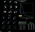
Optical coherence tomography (OCT) is a technique that displays images of the tissue by using the backscattered light.[citation needed] Not only conserving...
11 KB (1,669 words) - 23:39, 13 April 2025
Narrow-band imaging is an imaging technique for endoscopic diagnostic medical tests, where light of specific blue and green wavelengths is used to enhance...
5 KB (415 words) - 15:59, 27 April 2024

diffusion tensor imaging (DTI), has been used extensively to map white matter tractography in the brain. In diffusion weighted imaging (DWI), the intensity...
64 KB (9,251 words) - 19:41, 2 May 2025

for screening and diagnosis include optical coherence tomography (OCT), which can generate high-resolution images of the esophagus with moderate sensitivity...
43 KB (4,488 words) - 07:27, 31 May 2025
field diffraction patterns. Similar to optical coherence domain reflectometry (OCDR) and optical coherence tomography (OCT), a/LCI uses a broadband light...
15 KB (1,779 words) - 08:47, 28 May 2025
dilated fundus examination multifocal electroretinography (mfERG) optical coherence tomography (OCT) visual field test polysomnography pulmonary pletysmography...
29 KB (1,145 words) - 18:20, 23 May 2025

determine areas of dysplasia. Other gastroscopic modalities such as optical coherence tomography are being tested investigationally for similar applications....
77 KB (7,943 words) - 19:35, 16 June 2025
Endomicroscopy (category Medical imaging)
microscopy, although multi-photon microscopy and optical coherence tomography have also been adapted for endoscopic use. Commercially available clinical and pre-clinical...
12 KB (1,703 words) - 21:18, 26 May 2025
Kymograph (category Medical imaging)
three dimensions by incorporating depth-resolved imaging techniques such as optical coherence tomography. In this method, vertical sections of tissue (commonly...
6 KB (764 words) - 07:17, 10 April 2025

mucosa include confocal microscopy, magnification endoscopy and optical coherence tomography.[citation needed] Wong Kee Song, L. M.; Wong Kee Song, D. G.;...
9 KB (1,101 words) - 12:23, 30 July 2024

resonance imaging) can help in diagnosis. X-rays can determine the location and size of the narrowed airway portion. Optical coherence tomography (OCT) can...
11 KB (1,286 words) - 22:27, 23 May 2025

laser mammography medical imaging microscopy ophthalmology (includes Lasik and laser photocoagulation) optical coherence tomography optogenetics prostatectomy...
14 KB (1,402 words) - 23:37, 1 May 2025

DMSA scan (category 2D nuclear medical imaging)
injected into a patient, followed by imaging with a gamma camera after 2-3 hours. A DMSA scan is usually static imaging, while other radiotracers like DTPA...
4 KB (421 words) - 05:51, 12 September 2024

F; Dumas S; Denis P (2009). "Ultrasound biomicroscopy and optical coherence tomography imaging of filtering blebs after deep sclerectomy with new collagen...
30 KB (3,148 words) - 19:41, 24 May 2025
Gastric emptying study (category 2D nuclear medical imaging)
time-activity curve is produced from geometric mean of anterior and posterior imaging. Half-emptying time, the lag-phase duration for solid studies, and percentage...
4 KB (389 words) - 05:51, 12 September 2024

receiver and away from the x-rays source. The patients chin rests on the image receiver, which tilts the head up allowing the orbits to be clear of the...
3 KB (301 words) - 20:30, 14 January 2021

also known as lacrimal scintigraphy, is a nuclear medicine technique for imaging the lacrimal apparatus. It is used to identify obstructions, for example...
6 KB (574 words) - 20:09, 3 September 2024
intraocular pressure is around 16 mmHg [27]. Spectral domain optical coherence tomography can be used to view collector channel diameter change, collapse...
21 KB (3,131 words) - 00:18, 11 April 2025
computed tomographic MeSH E01.370.350.350.810.800 – tomography, spiral computed MeSH E01.370.350.400 – imaging, three-dimensional MeSH E01.370.350.400.200 –...
72 KB (6,253 words) - 16:46, 9 February 2024

(2004-08-16). "A Two-Axis Electrothermal Micromirror for Endoscopic Optical Coherence Tomography". IEEE Journal of Selected Topics in Quantum Electronics...
35 KB (3,691 words) - 06:42, 30 September 2024