
A micrograph is an image, captured photographically or digitally, taken through a microscope or similar device to show a magnified image of an object....
10 KB (954 words) - 20:30, 9 June 2025

Transmission electron microscopy (redirect from Transmission Electron Micrograph)
Transmission electron micrograph of dislocations in steel, which are faults in the structure of the crystal lattice at the atomic scale...
118 KB (15,040 words) - 22:12, 7 June 2025

Scanning electron microscope (redirect from Scanning electron micrograph)
and map their distribution. Due to the very narrow electron beam, SEM micrographs have a large depth of field yielding a characteristic three-dimensional...
68 KB (8,229 words) - 11:38, 16 May 2025

immature male analog, the immature glans penis. Micrograph of the primordial phallus, H&E stain. Micrograph of the primordial phallus, H&E stain. Aphallia...
1 KB (69 words) - 06:51, 17 March 2024

diagnosed incidentally by pathology. Micrograph of pinworms in the appendix, H&E stain High magnification micrograph of a pinworm in cross section in the...
25 KB (2,054 words) - 05:10, 2 June 2025

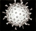
surface projections (the "peplomers" or "spikes"), which in electron micrographs of spherical particles create an image reminiscent of the solar corona...
11 KB (1,081 words) - 05:07, 2 June 2025

ruptured at the margin at the left in the image Micrograph of a placental infection (CMV placentitis) Micrograph of CMV placentitis A 3D Power Doppler image...
43 KB (4,622 words) - 00:36, 26 May 2025

Magneto-optic Kerr effect (redirect from Kerr micrograph)
In physics the magneto-optic Kerr effect (MOKE) or the surface magneto-optic Kerr effect (SMOKE) is one of the magneto-optic effects. It describes the...
11 KB (1,329 words) - 14:09, 30 April 2025

A fluorescence microscope is an optical microscope that uses fluorescence instead of, or in addition to, scattering, reflection, and attenuation or absorption...
25 KB (2,725 words) - 05:14, 1 April 2025
magnification micrograph of a granulosa cell tumour. H&E stain. High magnification micrograph of a thecoma. H&E stain. Low magnification micrograph of a thecoma...
14 KB (1,434 words) - 18:07, 5 January 2024

cystic carcinoma of the salivary gland, immunostain for S-100 protein Micrograph of adenoid cystic carcinoma, fine needle aspiration specimen, Pap stain...
13 KB (1,533 words) - 19:39, 18 September 2024
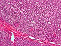
adenocarcinoma from biopsy. H&E stain. Micrograph of decidualized endometrium due to exogenous progesterone. H&E stain. Micrograph of decidualized endometrium due...
28 KB (3,140 words) - 04:42, 2 June 2025

Phase-contrast microscopy (redirect from Phase contrast micrograph)
Phase-contrast microscopy (PCM) is an optical microscopy technique that converts phase shifts in light passing through a transparent specimen to brightness...
12 KB (1,199 words) - 00:39, 31 May 2025

needed] Micrograph of a Kaposi sarcoma showing the characteristic spindle cells, high vascularity, and intracellular hyaline globs. H&E stain. Micrograph of...
37 KB (4,058 words) - 00:40, 26 May 2025
was discovered in 1973 by Ruth Bishop and her colleagues by electron micrograph images and accounts for approximately one third of hospitalisations for...
88 KB (10,052 words) - 05:25, 2 June 2025

needed] Low magnification micrograph of an SSL. Intermediate magnification micrograph of an SSL. High magnification micrograph of a SSL showing crypt branching...
8 KB (710 words) - 01:52, 5 February 2024

Transmission electron micrograph of multiple bacteriophages attached to a bacterial cell wall...
155 KB (18,324 words) - 19:17, 13 June 2025

magnification micrograph of a molluscum contagiosum lesion Low-magnification micrograph of molluscum contagiosum, H&E stain High-magnification micrograph of molluscum...
26 KB (2,857 words) - 13:13, 25 May 2025
club-shaped spikes that project from their surface, which in electron micrographs create an image reminiscent of the stellar corona, from which their name...
96 KB (10,611 words) - 20:30, 27 May 2025

5 cm long Intermediate magnification micrograph of a Leydig cell tumour, H&E stain High magnification micrograph of a Leydig cell tumour, H&E stain Cross-section...
15 KB (1,592 words) - 05:28, 28 May 2025

jpg". H&E stain. Intermediate magnification micrograph of a Warthin tumor. High magnification micrograph of a Warthin tumor showing the characteristic...
7 KB (686 words) - 19:32, 3 February 2025


analysis of the pathogenesis of MDBs. Micrograph showing a Mallory body. Original magnification 400X. H&E stain. Micrograph showing a Mallory body. Original...
5 KB (484 words) - 00:01, 30 October 2024

Electron micrograph of antenna surface detail of a wasp (Vespula vulgaris)...
20 KB (2,140 words) - 20:13, 6 June 2025

2012 A. wuliandei Zhu et al. 2020 Micrograph of actinomycosis, H&E stain Micrograph of actinomycosis, GMS stain Micrograph of actinomycosis, Gram stain Harz...
27 KB (1,377 words) - 06:35, 13 June 2025
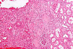
Micrograph of a renal oncocytoma. H&E stain. Micrograph of a renal oncocytoma. H&E stain. Micrograph of a renal oncocytoma. H&E stain. Micrograph of...
6 KB (438 words) - 19:03, 7 November 2023
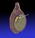
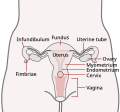
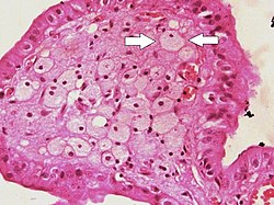
Vertical section of the testis, to show the arrangement of the ducts. Micrograph of the rete testis involved by seminoma. H&E stain. Tubular ectasia of...
5 KB (417 words) - 09:51, 5 August 2024

Medium-power magnification micrograph of a H&E stained slide showing a portion of a vaginal wall. Stratified squamous epithelium and underling connective...
172 KB (17,712 words) - 22:29, 7 June 2025
of the gallbladder). Micrograph of cholesterolosis of the gallbladder Micrograph of cholesterolosis of the gallbladder Micrograph of cholesterolosis of...
4 KB (232 words) - 08:41, 25 May 2025
Animated confocal micrograph of part of a biological neural network in a mouse's striatum...
8 KB (802 words) - 20:41, 9 June 2025