
Immune electron microscopy (more often called immunoelectron microscopy) is the equivalent of immunofluorescence, but it uses electron microscopy rather...
12 KB (1,417 words) - 01:10, 26 May 2025

Environmental scanning electron microscope (ESEM) Immune electron microscopy In situ electron microscopy Low-energy electron microscopy Microscope image processing...
48 KB (5,271 words) - 05:57, 20 June 2025
named coronavirus,: 96 due to their crown-like appearance. Her immune electron microscopy (IEM) innovations and insights contributed to research related...
24 KB (2,505 words) - 16:28, 26 May 2025

obtain X-ray diffraction results. In response to this, cryogenic electron microscopy (cryo-EM) emerged as a new, alternative method for studying virus...
32 KB (3,394 words) - 14:56, 23 May 2025

his colleagues isolated the Norwalk virus from volunteers using immune electron microscopy, a process that involves looking directly at antibody-antigen...
8 KB (738 words) - 13:57, 8 June 2025

Virology (section Electron microscopy)
The first images of viruses were obtained upon the invention of electron microscopy in 1931 by the German engineers Ernst Ruska and Max Knoll. In 1935...
59 KB (7,216 words) - 12:09, 24 June 2025
stretches and globular domains, as shown via high-resolution scanning electron microscopy. After stimulation of the neutrophil response, neutrophils lose their...
12 KB (1,478 words) - 21:39, 13 July 2024
antibody (peroxidase produces a dark brown color).[citation needed] Electron microscopy is a method that can take a picture of a whole virus and can reveal...
11 KB (1,346 words) - 12:41, 4 January 2024

Neurovascular unit (section Electron microscopy)
synapses and organelles. Scanning electron microscopy, on the other hand, provides 3D information by scanning a focused electron beam across the sample's surface...
38 KB (4,372 words) - 16:10, 6 January 2025
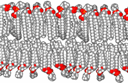
Lipid bilayer (section Electron microscopy)
on bilayers often require advanced techniques like electron microscopy and atomic force microscopy. When phospholipids are exposed to water, they self-assemble...
85 KB (10,132 words) - 08:56, 24 June 2025

classical complement pathway (C1q or C4) are usually not seen. Electron microscopy confirms electron-dense deposits in the mesangium that may extend to the subendothelial...
34 KB (3,950 words) - 22:38, 15 June 2025

mammalian immune system. A DC's main function is to process antigen material and present it on the cell surface to the T cells of the immune system. They...
35 KB (4,109 words) - 03:07, 22 June 2025

Transmission electron microscopy DNA sequencing is a single-molecule sequencing technology that uses transmission electron microscopy techniques. The method...
28 KB (3,145 words) - 04:04, 9 June 2025

children. Although no changes may be visible by light microscopy, changes on electron microscopy within the glomeruli may show a fusion of the foot processes...
21 KB (2,227 words) - 07:34, 9 May 2025

Lymphocyte (category Immune system)
(leukocyte) in the immune system of most vertebrates. Lymphocytes include T cells (for cell-mediated and cytotoxic adaptive immunity), B cells (for humoral...
23 KB (2,489 words) - 15:01, 8 June 2025

Clinical Skin Samples Using Scanning Electron Microscopy". In Janecek, Milos; Kral, Robert (eds.). Modern Electron Microscopy in Physical and Life Sciences....
13 KB (1,490 words) - 11:21, 3 April 2025
Fluorescence imaging (section Types of microscopy)
organism. Images can be produced from a variety of methods including: microscopy, imaging probes, and spectroscopy. Fluorescence itself, is a form of luminescence...
16 KB (1,896 words) - 20:52, 29 May 2025

Medical microbiology (section Microscopy)
electron microscopy are scanning electron microscopy and transmission electron microscopy. Transmission electron microscopy passes electrons through a...
38 KB (4,347 words) - 05:13, 26 May 2025

Sherman MB, Tsuprun VL (1990). "Negative staining of proteins". Electron Microscopy Reviews. 3 (1): 43–72. doi:10.1016/0892-0354(90)90013-I. PMID 1715774...
155 KB (18,324 words) - 19:17, 13 June 2025

stain, the GBM appears to have a "spiked" or "holey" appearance. On electron microscopy, subepithelial deposits that nestle against the glomerular basement...
22 KB (2,297 words) - 14:06, 24 June 2025

rotaviruses. Norwalk virus had been discovered by Albert Kapikian using immune electron microscopy and Ruth Bishop and colleagues had seen different particles that...
20 KB (2,270 words) - 03:28, 28 April 2025

single-wall carbon nanotubes, using transmission electron microscopy and scanning electron microscopy respectively, coupled with energy dispersive X-ray...
202 KB (21,775 words) - 02:38, 19 June 2025
proximal tubule of the nephron. Light microscopy findings in LCPT include proximal tubular swelling with electron microscopy findings showing proximal tubule...
14 KB (1,590 words) - 21:26, 19 February 2024
genetic recombination of its pili and surface proteins that interact with the immune system. Sexual transmission is through vaginal, anal, or oral sex. Sexual...
71 KB (8,032 words) - 12:14, 9 June 2025

fluorescence microscopy with imaging by focusing light and snap shooting instances to form a 3-D image. Transmission electron microscopy: Involves metal...
42 KB (5,255 words) - 08:55, 25 May 2025
peripheral zones on transmission electron microscopy is probably simplistic and may be in part a preparation artifact. Electron tomography with three-dimensional...
16 KB (1,941 words) - 15:47, 25 May 2025

Infection (section Microscopy)
agent. Microscopy may be carried out with simple instruments, such as the compound light microscope, or with instruments as complex as an electron microscope...
118 KB (12,873 words) - 15:07, 23 May 2025

century, electron microscopy also saw a drastic revolution with the development of more coherent electron sources, aberration correction for electron microscopes...
20 KB (2,191 words) - 00:09, 18 June 2025
in a granular fluorescent staining pattern. Electron microscopy reveals electron-dense subendothelial immune complexes (between endothelium and basement...
23 KB (2,439 words) - 14:57, 23 May 2025
aspect ratio structure called CuHARS. Scanning electron microscopy (SEM) and transmission electron microscopy (TEM) of CuHARS revealed linear morphology and...
10 KB (1,226 words) - 13:41, 19 April 2025