 | The vertebrate cerebrum (brain) is formed by two cerebral hemispheres that are separated by a groove, the longitudinal fissure. The brain can thus be described... 13 KB (1,575 words) - 21:39, 14 February 2024 |
 | Lateralization of brain function (redirect from Cerebral hemispheric dominance) brain into two distinct cerebral hemispheres, connected by the corpus callosum. Although the macrostructure of the two hemispheres appears to be almost identical... 39 KB (4,316 words) - 16:31, 8 April 2024 |
 | Cerebrum (section Cerebral hemispheres) endbrain is the largest part of the brain containing the cerebral cortex (of the two cerebral hemispheres), as well as several subcortical structures, including... 16 KB (1,779 words) - 08:22, 3 January 2024 |
 | radiograms. Areas supplied by the middle cerebral artery include: The bulk of the lateral surface of the hemisphere; except for the superior inch of the frontal... 11 KB (1,289 words) - 08:08, 13 April 2024 |
| defects more precisely. Middle cerebral artery lesions mostly affect the dominant hemisphere i.e. the left cerebral hemisphere. Hemiparesis or hemiplegia... 5 KB (488 words) - 04:13, 21 February 2023 |
 | Gyrus (redirect from Cerebral gyri) gyri) is a congenital malformation of the cerebral hemisphere, resulting in unusually thick gyri in the cerebral cortex. Pachygyria is used to describe brain... 8 KB (845 words) - 17:38, 26 April 2024 |
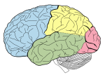
 | Lobes of the brain (redirect from Cerebral lobes) identifiable zones of the human cerebral cortex, and they comprise the surface of each hemisphere of the cerebrum. The two hemispheres are roughly symmetrical... 15 KB (1,722 words) - 17:31, 23 February 2024 |
Northern celestial hemisphere Southern celestial hemisphere A cultural hemisphere The near or far side of the Moon A cerebral hemisphere, a division of the... 1 KB (176 words) - 06:45, 7 November 2023 |
 | white matter structure situated in the inferomedial part of each cerebral hemisphere of the brain. It carries information past the basal ganglia, separating... 11 KB (1,261 words) - 19:12, 20 June 2023 |
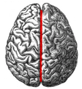
 | right parts by the longitudinal fissure, which separates the two cerebral hemispheres that are joined beneath the cortex by the corpus callosum. In most... 67 KB (7,791 words) - 08:13, 30 April 2024 |
 | cortical region of the limbic system, on the medial surface of each cerebral hemisphere of the mammalian brain, consisting of parts of the frontal, parietal... 5 KB (491 words) - 16:00, 7 December 2023 |
 | Falx cerebri (redirect from Cerebral falx) cerebral falx) is a large, crescent-shaped fold of dura mater that descends vertically into the longitudinal fissure between the cerebral hemispheres... 8 KB (927 words) - 16:17, 18 January 2024 |
| Sulcus (neuroanatomy) (redirect from Cerebral sulci) sulcus (Latin: "furrow"; pl.: sulci) is a depression or groove in the cerebral cortex. It surrounds a gyrus (pl. gyri), creating the characteristic folded... 9 KB (1,061 words) - 16:28, 13 April 2024 |
| lobes of the brain in mammals, and is located at the front of each cerebral hemisphere (in front of the parietal lobe and the temporal lobe). It is parted... 22 KB (2,687 words) - 14:38, 24 April 2024 |
 | Longitudinal fissure (redirect from Longitudinal cerebral fissure) interhemispheric fissure) is the deep groove that separates the two cerebral hemispheres of the vertebrate brain. Lying within it is a continuation of the... 19 KB (2,348 words) - 16:39, 6 April 2024 |
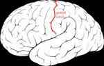 | video. Demonstrating position of the central sulcus of the left cerebral hemisphere Primary motor cortex Primary somatosensory cortex Luigi Rolando List... 17 KB (1,989 words) - 15:55, 19 February 2024 |
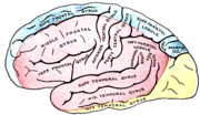
| Temporal lobe (section Dominant hemisphere) lobes of the cerebral cortex in the brain of mammals. The temporal lobe is located beneath the lateral fissure on both cerebral hemispheres of the mammalian... 15 KB (1,517 words) - 22:50, 12 December 2023 |
 | Animation. Parahippocampal gyrus shown red. Medial surface of left cerebral hemisphere. Parahippocampal gyrus shown in orange. Human brain inferior-medial... 8 KB (781 words) - 10:52, 29 April 2023 |
| They are structurally isolated in their respective cerebral hemispheres by the separation of the cerebral fissure. At the front edge of the occipital lobe... 11 KB (1,315 words) - 14:37, 3 April 2024 |
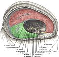
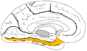
| supply. Medial surface of cerebral hemisphere, showing areas supplied by cerebral arteries. Areas supplied by the posterior cerebral artery shown in yellow... 12 KB (1,216 words) - 10:50, 30 October 2023 |
 | Fusiform gyrus seen in a ventral view Fusiform gyrus, in the right cerebral hemisphere. 3D view of the fusiform gyrus. Yellow: temporal section, red: occipito-temporal... 18 KB (1,966 words) - 14:44, 3 April 2024 |
 | Wernicke's area (category Cerebral cortex) located in the superior temporal gyrus in the dominant cerebral hemisphere, which is the left hemisphere in about 95% of right-handed individuals and 70% of... 23 KB (2,748 words) - 22:06, 4 March 2024 |
 | experimental conditions. Lateral surface of left cerebral hemisphere, viewed from above. Left cerebral hemisphere, viewed from the back. (Intraparietal sulcus... 9 KB (880 words) - 06:47, 9 April 2024 |
| The anterior cerebral artery (ACA) is one of a pair of cerebral arteries that supplies oxygenated blood to most midline portions of the frontal lobes and... 10 KB (1,058 words) - 10:17, 8 November 2023 |
 | consists of two cerebral hemispheres. Each hemisphere has an inner core composed of white matter, and an outer surface – the cerebral cortex – composed... 169 KB (18,798 words) - 18:30, 26 April 2024 |
 | Association fiber (category Cerebral white matter) Association fibers are axons that connect cortical areas within the same cerebral hemisphere. In human neuroanatomy, axons (nerve fibers) within the brain, can... 4 KB (321 words) - 16:03, 22 January 2024 |
 | Precuneus (category Medial surface of cerebral hemisphere) in 1879. The precuneus is located on the inside between the two cerebral hemispheres in the rear region between the somatosensory cortex and forward of... 23 KB (2,675 words) - 17:33, 18 February 2024 |
whereas focal atrophy affects cells in a specific location. If the cerebral hemispheres (the two lobes of the brain that form the cerebrum) are affected... 20 KB (2,146 words) - 15:05, 20 December 2023 |
| The parietal lobe is one of the four major lobes of the cerebral cortex in the brain of mammals. The parietal lobe is positioned above the temporal lobe... 20 KB (2,238 words) - 19:38, 8 April 2024 |
 | Cuneus (category Medial surface of cerebral hemisphere) left cerebral hemisphere. Medial surface of left cerebral hemisphere. Cuneus is visible at left in green. Infero-medial surface of right cerebral hemisphere... 3 KB (340 words) - 16:08, 3 January 2024 |