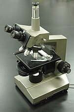
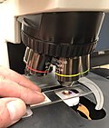
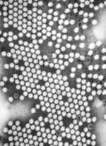
• Thumbnail for Dark-field microscopy
    Dark-field microscopy (also called dark-ground microscopy) describes microscopy methods, in both light and electron microscopy, which exclude the unscattered...
    11 KB (1,226 words) - 18:15, 15 September 2023
  • Thumbnail for Weak-beam dark-field microscopy
    Weak beam dark field (WBDF) microscopy is a type of transmission electron microscopy (TEM) dark field imaging technique that allows for the visualization...
    23 KB (3,407 words) - 16:10, 17 October 2023
  • Thumbnail for Bright-field microscopy
    appearance of a bright-field microscopy image is a dark sample on a bright background, hence the name. The light path of a bright-field microscope is extremely...
    6 KB (672 words) - 01:31, 4 December 2023
  • Thumbnail for Annular dark-field imaging
    Transmission electron microscopy Scanning transmission electron microscopy Dark field microscopy Otten, Max T. (1992). "High-Angle annular dark-field imaging on...
    6 KB (760 words) - 08:25, 8 November 2023
  • Dark-field X-ray microscopy (DFXM or DFXRM) is an imaging technique used for multiscale structural characterisation. It is capable of mapping deeply embedded...
    36 KB (3,801 words) - 16:16, 11 March 2024
  • Thumbnail for Microscopy
    Microscopy is the technical field of using microscopes to view objects and areas of objects that cannot be seen with the naked eye (objects that are not...
    69 KB (8,317 words) - 13:29, 18 April 2024
  • Thumbnail for Metallography
    in some cases, may provide superior images with greater detail. Dark-field microscopy (DF), is an alternative method of observation that provides high-contrast...
    19 KB (2,373 words) - 09:07, 5 December 2023
  • Thumbnail for Light sheet fluorescence microscopy
    fluorescence microscopy, which has already been used successfully for dark field microscopy under the name ultramicroscopy. In this type of microscopy, the illumination...
    41 KB (4,599 words) - 02:00, 26 January 2024
  • Hemaview or nutritional blood analysis is the use of high-resolution dark field microscopy to observe live blood cells. Live blood analysis is promoted by...
    18 KB (1,907 words) - 17:22, 27 February 2024
  • Field-emission microscopy (FEM) is an analytical technique that is used in materials science to study the surfaces of needle apexes. The FEM was invented...
    6 KB (741 words) - 23:18, 12 August 2023
  • Thumbnail for Syphilis
    made by using blood tests; the bacteria can also be detected using dark field microscopy. The Centers for Disease Control and Prevention (U.S.) recommend...
    98 KB (10,427 words) - 12:21, 21 April 2024
  • dark field microscopy The Dark Fields , a 2001 novel by Alan Glynn Dark Fields (2006 film), a horror film directed by Mark McNabb and Al Randall Dark Fields...
    735 bytes (124 words) - 19:15, 21 December 2015
  • Thumbnail for Phase-contrast microscopy
    Phase-contrast microscopy is particularly important in biology. It reveals many cellular structures that are invisible with a bright-field microscope, as...
    12 KB (1,179 words) - 17:23, 28 March 2024
  • spirochetes are difficult to Gram-stain but may be visualized using dark field microscopy or Warthin–Starry stain. Examples include: Leptospira species, which...
    4 KB (305 words) - 13:10, 18 December 2023
  • Thumbnail for Borrelia
    pallidum. This bacterium must be viewed using dark-field microscopy, which make the cells appear white against a dark background. Borrelia species are grown...
    28 KB (2,215 words) - 02:44, 29 January 2024
  • Thumbnail for Localized surface plasmon
    surface plasmons are dark-field microscopy, UV-vis-NIR spectroscopy, and surface-enhanced Raman scattering (SERS). With dark-field microscopy, it is possible...
    12 KB (1,370 words) - 06:40, 7 November 2023
  • Thumbnail for Leptospirosis
    of its movement. The bacteria are traditionally visualised using dark-field microscopy without staining. A total of 66 species of Leptospira has been identified...
    76 KB (8,120 words) - 19:29, 3 March 2024
  • Thumbnail for Histology
    film. Individual silver grains in the film are visualized with dark field microscopy. Recently, antibodies have been used to specifically visualize proteins...
    33 KB (3,169 words) - 21:39, 6 March 2024
  • Thumbnail for Nonvenereal endemic syphilis
    patient as well as laboratory testing of material from the lesions (dark-field microscopy). The responsible spirochaete is readily identifiable on sight in...
    4 KB (371 words) - 19:01, 3 December 2023
  • Thumbnail for Transmission electron microscopy
    Transmission electron microscopy (TEM) is a microscopy technique in which a beam of electrons is transmitted through a specimen to form an image. The specimen...
    117 KB (15,027 words) - 18:11, 19 April 2024
  • near-field (photon-tunneling microscopy as well as those that use the Pendry Superlens and near field scanning optical microscopy) or on the far-field. Among...
    87 KB (10,121 words) - 08:36, 16 April 2024
  • and TPHA, will be positive, and the spirochetes can be seen on dark field microscopy of samples taken from the early papules.[citation needed] The disease...
    4 KB (395 words) - 23:24, 1 February 2024
  • Thumbnail for Stereo microscope
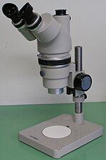
    illuminators can be used for dark field microscopy, using either reflected or transmitted light. Great working distance and depth of field are important qualities...
    11 KB (1,406 words) - 08:10, 28 September 2023
  • Thumbnail for Total internal reflection microscopy
    reflection microscopy is a specialized optical imaging technique for object tracking and detection utilizing the light scattered from an evanescent field in the...
    5 KB (684 words) - 00:56, 4 December 2023
  • Thumbnail for Microorganism
    his own design. Robert Hooke, a contemporary of Leeuwenhoek, also used microscopy to observe microbial life in the form of the fruiting bodies of moulds...
    73 KB (7,720 words) - 14:19, 4 March 2024
  • Negative stain (category Electron microscopy stains)
    positive staining, in which the actual specimen is stained. For bright-field microscopy, negative staining is typically performed using a black ink fluid such...
    6 KB (601 words) - 02:00, 4 December 2021
  • Thumbnail for Differential interference contrast microscopy
    interference contrast (DIC) microscopy, also known as Nomarski interference contrast (NIC) or Nomarski microscopy, is an optical microscopy technique used to enhance...
    13 KB (1,567 words) - 05:26, 19 May 2023
  • Thumbnail for Photoacoustic microscopy
    conventional microscopy methods. Furthermore, photoacoustic microscopy is especially useful in the field of biomedical imaging due to its scalability. By adjusting...
    16 KB (2,065 words) - 17:21, 29 September 2023
  • Thumbnail for Optical microscope
    illumination. For illumination techniques like dark field, phase contrast and differential interference contrast microscopy additional optical components must be...
    52 KB (5,972 words) - 03:47, 11 April 2024
  • is swapped for one inside it: this has long been implemented in dark-field microscopy. Nor are information-theoretical rules broken when superimposing...
    28 KB (3,037 words) - 02:54, 20 March 2024