
The infundibulum (also known as conus arteriosus) is a conical pouch formed from the upper and left angle of the right ventricle in the chordate heart, from...
1 KB (147 words) - 04:04, 22 March 2024
Hair follicle: the infundibulum is the cup or funnel in which a hair follicle grows. Infundibulum (heart): The infundibulum of the heart, or conus arteriosus...
2 KB (360 words) - 02:12, 21 November 2020
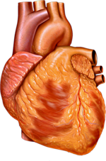
The heart is a muscular organ found in most animals. This organ pumps blood through the blood vessels of the circulatory system. The pumped blood carries...
143 KB (16,874 words) - 00:43, 25 September 2024

Cardiac conduction system (redirect from Heart conduction system)
conduction system of the heart) transmits the signals generated by the sinoatrial node – the heart's pacemaker, to cause the heart muscle to contract, and...
16 KB (1,840 words) - 02:31, 30 January 2024

Cardiac surgery (redirect from Heart surgery)
stenosis. Later that year, he designed a punch to resect a stenosed infundibulum, which is often associated with Tetralogy of Fallot. Many thousands of...
30 KB (3,223 words) - 22:49, 30 August 2024

Aortic valve (redirect from Aortic heart valve)
valve in the heart of humans and most other animals, located between the left ventricle and the aorta. It is one of the four valves of the heart and one of...
12 KB (1,504 words) - 12:27, 17 August 2024
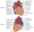
two upper chambers in the heart that receives blood from the circulatory system. The blood in the atria is pumped into the heart ventricles through the atrioventricular...
24 KB (2,579 words) - 07:07, 16 August 2024

the heart valves closing in a healthy 16 year old girl. The stethoscope is at the tricuspid area. Problems playing this file? See media help. A heart valve...
22 KB (2,640 words) - 19:32, 16 August 2024
ventricle is one of two large chambers located toward the bottom of the heart that collect and expel blood towards the peripheral beds within the body...
20 KB (2,368 words) - 12:32, 31 July 2024

Chordae tendineae (redirect from Heart strings)
tendineae (sg.: chorda tendinea) or tendinous cords, colloquially known as the heart strings, are inelastic cords of fibrous connective tissue that connect the...
8 KB (797 words) - 20:41, 26 August 2024

horseshoe-shaped area expands to form the future ventricular infundibulum and the ventricular regions, as the heart tube continues to expand. The tube starts receiving...
24 KB (3,015 words) - 18:56, 20 April 2024

Circulatory system (redirect from Three chambered heart)
The circulatory system is a system of organs that includes the heart, blood vessels, and blood which is circulated throughout the entire body of a human...
50 KB (5,592 words) - 00:50, 25 September 2024
terminal sulcus is a groove on the outer surface of the right atrium of the heart marking the transition between the sinus venarum cavarum (which has a distinct...
2 KB (201 words) - 15:40, 14 February 2024
Mitral valve (redirect from Mitral heart valve)
one of the four heart valves. It has two cusps or flaps and lies between the left atrium and the left ventricle of the heart. The heart valves are all...
20 KB (2,337 words) - 21:31, 18 July 2024
Cardiac skeleton (redirect from Fibrous skeleton of the heart)
fibrous skeleton of the heart, is a high-density homogeneous structure of connective tissue that forms and anchors the valves of the heart, and influences the...
13 KB (1,529 words) - 17:26, 6 July 2024
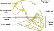
Purkinje fibers (redirect from Purkinje cell heart)
subendocardial branches) are located in the inner ventricular walls of the heart, just beneath the endocardium in a space called the subendocardium. The...
8 KB (839 words) - 23:06, 31 August 2024
The left border of heart (or obtuse margin) is formed from the rounded lateral wall of the left ventricle. It is called the 'obtuse' margin because of...
1 KB (192 words) - 21:38, 15 May 2024

Pericardium (redirect from Heart sac)
pericardia), also called pericardial sac, is a double-walled sac containing the heart and the roots of the great vessels. It has two layers, an outer layer made...
13 KB (1,465 words) - 13:37, 20 July 2024
The right border of the heart (right margin of heart) is a long border on the surface of the heart, and is formed by the right atrium. The atrial portion...
970 bytes (114 words) - 11:54, 18 August 2019
Pulmonary valve (redirect from Pulmonary heart valve)
situated at a superior level than the aortic orifice. At the apex of the infundibulum, the pulmonary orifice is guarded by three semilunar cusps - two anterior...
4 KB (444 words) - 01:26, 24 June 2024

Cardiac muscle (redirect from Heart muscle)
Cardiac muscle (also called heart muscle or myocardium) is one of three types of vertebrate muscle tissues, the others being skeletal muscle and smooth...
42 KB (5,130 words) - 05:03, 14 August 2024
vessels of the heart, and is deficient in front, where it is crossed by the root of the pulmonary trunk. On the posterior surface of the heart, the coronary...
6 KB (691 words) - 21:16, 8 April 2024

Pericardial fluid (section Ischemic heart disease)
each other with each heart beat. Ben-Horin et al. (2005) studied the composition of pericardial fluid in patients undergoing open heart surgery. They found...
5 KB (530 words) - 16:46, 29 July 2024
Coronary circulation (redirect from Blood supply of heart)
arteries and veins that supply the heart muscle (myocardium). Coronary arteries supply oxygenated blood to the heart muscle. Cardiac veins then drain away...
16 KB (1,977 words) - 21:49, 28 November 2023

The fossa ovalis is a depression in the right atrium of the heart, at the level of the interatrial septum, the wall between right and left atrium. The...
7 KB (907 words) - 15:30, 18 January 2024
of cardiac muscle (heart muscle) in all animals is initiated by electrical impulses known as action potentials that in the heart are known as cardiac...
11 KB (1,456 words) - 22:27, 11 August 2024

coronary circulation, which transport oxygenated blood to the heart muscle. The heart requires a continuous supply of oxygen to function and survive...
11 KB (1,265 words) - 07:56, 5 July 2024

of the heart, causing it to contract. In a healthy heart, the SA node continuously produces action potentials, setting the rhythm of the heart (sinus...
24 KB (2,805 words) - 08:57, 15 September 2024
connects the heart's atria and ventricles to coordinate beating in the top of the heart; it is part of the electrical conduction system of the heart. The AV...
11 KB (1,218 words) - 17:51, 24 April 2024
In the heart's conduction system, Bachmann's bundle (also called the Bachmann bundle or the interatrial band) is a branch of the anterior internodal tract...
4 KB (474 words) - 14:21, 18 May 2024