 | endocardium in a space called the subendocardium. The Purkinje fibers are specialized conducting fibers composed of electrically excitable cells. They are... 8 KB (839 words) - 02:21, 1 April 2024 |
 | parallel fibers from the deeper-layers pass. These parallel fibers make relatively weaker excitatory (glutamatergic) synapses to spines in the Purkinje cell... 29 KB (3,273 words) - 06:14, 8 April 2024 |
the cerebellum Purkinje fibers, located in the heart The visual Purkinje effect of how human beings do not see color in dim light Purkinje images, reflections... 606 bytes (111 words) - 19:04, 8 December 2021 |
connect with the Purkinje fibers. This passage takes approximately 25 ms. The Purkinje fibers are additional myocardial conductive fibers that spread the... 47 KB (5,784 words) - 11:54, 12 June 2023 |
| bundles and fascicles give rise to thin filaments known as Purkinje fibers. These fibers distribute the impulse to the ventricular muscle. The ventricular... 7 KB (772 words) - 00:43, 21 December 2023 |
 | His, and through the bundle branches to Purkinje fibers in the walls of the ventricles. The Purkinje fibers transmit the signals more rapidly to stimulate... 16 KB (1,840 words) - 02:31, 30 January 2024 |
 | Cerebellum (redirect from Purkinje layer) following pathway: mossy fibers → granule cells → parallel fibers → Purkinje cells → deep nuclei. Climbing fibers project to Purkinje cells and also send collaterals... 94 KB (11,557 words) - 13:28, 21 April 2024 |
| is the Bundle of His. The left and right bundle branches, and the Purkinje fibers, will also produce a spontaneous action potential at a rate of 30-40... 11 KB (1,469 words) - 21:35, 12 February 2024 |
 | transmitting cardiac action potentials from the bundle of His to the Purkinje fibers. There are two branches of the bundle of His: the left bundle branch... 4 KB (506 words) - 08:44, 11 December 2023 |
 | ventricles the signal is carried by specialized tissue called the Purkinje fibers which then transmit the electric charge to the heart muscle. A racing... 143 KB (16,888 words) - 19:48, 24 April 2024 |
 | potential (EPSP) in Purkinje cells. In this way climbing fibers (CFs) perform a central role in motor behaviors. The climbing fibers carry information from... 7 KB (689 words) - 15:59, 4 February 2022 |
 | bundle of His (AV bundle), down the bundle branches, and into the Purkinje fibers. This would depolarize the ventricles and cause them to contract. In... 6 KB (716 words) - 03:39, 20 April 2024 |
 | small size. 'U' waves are thought to represent repolarization of the Purkinje fibers. However, the exact source of the U wave remains unclear. The most... 7 KB (987 words) - 01:55, 16 December 2023 |
 | the atrioventricular node down into the bundle of His and into the Purkinje fibers, spreading down and to the left throughout the ventricles. This orderly... 89 KB (9,808 words) - 16:45, 21 April 2024 |
 | produces an action potential. Other cells within the heart (including the Purkinje fibers and atrioventricular node) can also initiate action potentials; however... 24 KB (2,806 words) - 17:50, 24 April 2024 |
 | the bundle of His, and the Purkinje fibers are larger in diameter and conduct signals at a fast rate. The Purkinje fibers rapidly conduct electrical signals;... 42 KB (5,108 words) - 13:34, 28 April 2024 |
 | Purkinje fibers (p-fibers) found in the hearts moderator bands are a specialized cardiac muscle fiber that causes the heart to contract. The Purkinje... 122 KB (13,267 words) - 14:54, 18 April 2024 |
 | Anatomy of the cerebellum (section Purkinje layer) nuclei in the brainstem. Each Purkinje cell receives excitatory input from 100,000 to 200,000 parallel fibers. Parallel fibers are said to be responsible... 32 KB (3,180 words) - 19:42, 25 April 2024 |
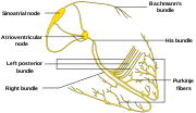
 | pacemaker SA node Bachmann's bundle AV node bundle of His bundle branches Purkinje fibers Pericardial cavity pericardial sinus Pericardium fibrous pericardium... 8 KB (785 words) - 10:58, 17 January 2024 |
 | signal travels into the Purkinje fibers. The division of the signal into a right and left bundle and then into the Purkinje fibers allows for a simultaneous... 15 KB (1,805 words) - 20:34, 30 October 2023 |
 | atrioventricular node and into the bundle of His, from which it travels along Purkinje fibers to reach and depolarize the ventricles. This sinus rhythm is important... 8 KB (890 words) - 11:29, 26 April 2024 |
 | atria and through the circuits known as the bundle of His and the Purkinje fibers—all which stimulate contractions of both ventricles. The programmed... 20 KB (1,882 words) - 12:31, 3 April 2024 |
completed. EADs most commonly originate in mid-myocardial cells and Purkinje fibers, but can develop in other cardiac cells that carry an action potential... 5 KB (546 words) - 01:14, 29 December 2023 |
conduct impulses through the interventricular septum and into the Purkinje fibers, as these are responsible for the depolarization of contractile cells... 81 KB (8,705 words) - 23:54, 13 March 2024 |
 | contraction (PVC) is a common event where the heartbeat is initiated by Purkinje fibers in the ventricles rather than by the sinoatrial node. PVCs may cause... 36 KB (3,827 words) - 18:10, 23 March 2024 |
component of the natural pacemaker. First described in the late 1970s in Purkinje fibers and sinoatrial myocytes, the cardiac pacemaker "funny" (If) current... 10 KB (1,195 words) - 21:33, 8 November 2023 |
 | the Purkinje fibers at the bottom (apex) of the heart, causing ventricular contraction.[citation needed] In addition to the SAN, the AVN and Purkinje fibres... 46 KB (5,430 words) - 18:10, 24 April 2024 |
| parallel fiber which innervates Purkinje cells. The vast majority of granule cell axonal synapses are found on the parallel fibers. The parallel fibers are... 27 KB (3,177 words) - 17:30, 26 April 2024 |
 | pacemaker SA node Bachmann's bundle AV node bundle of His bundle branches Purkinje fibers Pericardial cavity pericardial sinus Pericardium fibrous pericardium... 8 KB (798 words) - 15:26, 14 February 2024 |
 | pathways contain the sinoatrial node, the atrioventricular node, and the Purkinje fibers. (Exceptions such as accessory pathways may occur in this firewall... 20 KB (2,437 words) - 04:50, 20 April 2024 |