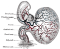
 | The vitelline veins are veins that drain blood from the yolk sac and the gut tube during gestation. They run upward at first in front, and subsequently... 6 KB (592 words) - 11:35, 15 February 2024 |
 | The vitelline arteries are the arterial counterpart to the vitelline veins. Like the veins, they play an important role in the vitelline circulation of... 1 KB (141 words) - 14:09, 31 October 2023 |
 | is formed of six paired veins, the vitelline veins, umbilical veins, and the cardinal veins. In the systemic circulation, veins serve to return oxygen-depleted... 49 KB (6,218 words) - 12:52, 5 May 2024 |
 | the hepatic veins are the veins that drain venous blood from the liver into the inferior vena cava (as opposed to the hepatic portal vein which conveys... 7 KB (755 words) - 23:46, 11 March 2024 |
 | vitelline arteries (a branch of the dorsal aorta), and after circulating through a wide-meshed capillary plexus, is returned by the vitelline veins to... 1 KB (160 words) - 05:00, 11 April 2024 |
of a human embryo Vitelline membrane, membrane surrounding an ovum Vitelline veins, veins that drain blood from the yolk sac Vitelline masked weaver (Ploceus... 982 bytes (149 words) - 17:24, 28 October 2016 |
| capillary plexus, is returned by the vitelline veins to the tubular heart of the embryo. This constitutes the vitelline circulation, which in humans serves... 8 KB (972 words) - 21:55, 3 May 2024 |
 | Circulatory system (redirect from Systemic Veins) human venous system develops mainly from the vitelline veins, the umbilical veins and the cardinal veins, all of which empty into the sinus venosus. About... 50 KB (5,591 words) - 06:54, 29 April 2024 |
 | the descending aorta. Each primitive aorta anteriorly receives the vitelline vein from the yolk-sac, and is prolonged[clarification needed] backward on... 2 KB (199 words) - 14:31, 14 August 2023 |
 | rest of their extent. It receives blood from the vitelline vein, umbilical vein and common cardinal vein.[citation needed] The sinus venosus originally... 3 KB (306 words) - 09:30, 5 January 2023 |
thymus. Blood islands have been seen in the area vasculosa in the vitelline veins and arteries, and in the dorsal aorta. Tortora, Gerard J.; Derrickson... 4 KB (392 words) - 18:00, 28 January 2023 |
 | pole receives blood from three major veins: the vitelline vein, the umbilical vein and the common cardinal vein. The sinus opening moves clockwise. This... 24 KB (3,015 words) - 18:56, 20 April 2024 |
 | liver sinusoids and empties into the central vein of each lobule. The central veins coalesce into hepatic veins, which leave the liver and drain into the... 85 KB (10,183 words) - 16:46, 1 May 2024 |
 | The esophagus also lies in front of parts of the hemiazygos veins and the intercostal veins on the right side. The vagus nerve divides and covers the esophagus... 46 KB (5,414 words) - 04:15, 5 May 2024 |
usually result from a developmental abnormality of the vitelline veins, which connect the portal vein to the caudal vena cava. Thus in the juvenile and adult... 12 KB (1,441 words) - 14:52, 20 November 2023 |
 | sac, which lies outside the zona pellucida (in mammals), known as the vitelline membrane in other animals. In insects, it is developed by the follicle... 11 KB (1,223 words) - 16:02, 18 January 2024 |
 | involution of the right umbilical vein leading to weakening of the body wall and gut herniation Disruption of the right vitelline (yolk sac) artery with subsequent... 16 KB (1,675 words) - 19:56, 11 April 2024 |
 | large intestine. In the fetus the ileum is connected to the navel by the vitelline duct. In roughly 2−4% of humans, this duct fails to close during the first... 13 KB (1,593 words) - 15:11, 2 May 2024 |
 | their final habitation, the inferior mesenteric veins. Individual females cannot enter the mesenteric veins. Sex organs, the gonads, are also incompletely... 57 KB (6,881 words) - 21:36, 17 February 2024 |
 | by a similarly named vein, the inferior mesenteric vein, which drains into the splenic vein. The IMV drains to the portal vein and does therefore not... 7 KB (696 words) - 23:28, 23 April 2024 |
umbilical vein, hematogenous spread, or via remnant structures such as the falciform ligament, median umbilical ligament, or a remnant of the vitelline duct... 5 KB (524 words) - 00:58, 12 December 2023 |
 | the splenic vein. Located under this portion of the superior mesenteric artery, between it and the aorta, are the following: left renal vein - travels between... 6 KB (563 words) - 22:38, 13 March 2024 |
 | the hepatic veins. In contrast to the drainage of midgut and hindgut structures by the superior mesenteric vein and inferior mesenteric vein respectively... 9 KB (852 words) - 16:41, 11 February 2024 |
 | venosus receives blood from the three major veins: the vitelline, the umbilical and the common cardinal veins. During the first two months of development... 42 KB (5,252 words) - 16:42, 1 May 2024 |
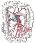
 | gastrointestinal tract. The sac is surrounded by a network of vitelline arteries and veins. Over time, these arteries consolidate into the three main arteries... 47 KB (4,982 words) - 22:56, 4 May 2024 |
 | layer can be seen in unfertilized eggs, because after fertilization, the vitelline, chitinous, and lipid layers form. In Canada in 1970, a postgraduate student... 10 KB (1,168 words) - 22:05, 22 April 2024 |
 | Amphibian egg: Jelly capsule Vitelline membrane Perivitelline fluid Yolk plug Embryo... 161 KB (17,759 words) - 19:10, 1 May 2024 |
umbilical arteries MeSH A16.254.789.807 – umbilical veins MeSH A16.254.835 – urachus MeSH A16.254.891 – vitelline duct MeSH A16.254.940 – Wolffian duct MeSH A16... 5 KB (424 words) - 16:46, 9 February 2024 |
 | markers onto chick veins (or vice versa), showcasing the plasticity of the system. Reversing flow patterns in arteries and/or veins can also have the same... 41 KB (4,873 words) - 01:31, 24 April 2024 |
 | male above, small wingless female below Tubular veins run through the two-layered membranous wing. Veins are connected to the haemocoel and in theory allow... 95 KB (10,293 words) - 03:58, 24 March 2024 |