
 French
 Deutsch
Entamoeba gingivalis
| Entamoeba gingivalis | |
|---|---|
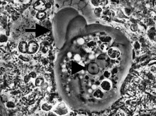 | |
| Entamoeba gingivalis from periodontal pocket [1] | |
| Scientific classification | |
| Domain: | Eukaryota |
| Phylum: | Amoebozoa |
| Family: | Entamoebidae |
| Genus: | Entamoeba |
| Species: | E. gingivalis |
| Binomial name | |
| Entamoeba gingivalis Gros, 1849 | |

Entamoeba gingivalis is an opportunistic Amoebozoa[citation needed] (reported by some as an effect of disease; not a cause [hence status as a commensal])[2][3][4] and is the first amoeba in humans to be described.
It is found in the mouth[5] inside the gingival pocket biofilm near the base of the teeth, and in periodontal pockets.[1] Entamoeba gingivalis is found in 95% of people with gum disease and rarely in people with healthy gums.[6][7] Cyst formation is not present; therefore transmission is direct from one person to another by kissing, or by sharing eating utensils. Only the trophozoites are formed and the size is usually 20 micrometers to 150 micrometers in diameter. Entamoeba gingivalis have pseudopodia that allow them to move quickly and phagocytise the nucleus of polynuclear neutrophils by exonucleophagy in periodontal disease.[8] Their spheroid nucleus is 2 micrometers to 4 micrometers in diameter and contains a small central endosome. There are numerous food vacuoles, which consists mostly of phagocitised PMN nucleus, blood cells, and bacteria. It also causes pyorrhoea.
Media[edit]
The main activity of the amoeba Entamoeba gingivalis in the infected periodontal crevices, besides moving, consists in feeding on the nucleus of white blood cells. The amoeba penetrates into the cytoplasm to reach the nucleus and literally suctions its contents via the negative pressure of the pseudopod. The food so gulped down is gradually digested inside the endoplasm. Phagocytosis can sometimes continue for more than 20 polynuclear neutrophil nuclei. This activity leaves a denucleated cell, unable to achieve either its NETS activity or its preprogrammed apoptosis. It will release PMN-uncontrolled proteolytic enzymes on surrounding tissues and could be considered a pathogen from this vampirising activity.
References[edit]
- ^ a b Bonner M, Amard V, Bar-Pinatel C, Charpentier F, Chatard JM, Desmuyck Y, et al. (2014). "Detection of the amoeba Entamoeba gingivalis in periodontal pockets". Parasite. 21: 30. doi:10.1051/parasite/2014029. PMC 4077299. PMID 24983705.
- ^ Jian B, Kolansky AS, Baloach ZW, Gupta PK (September 2008). "Entamoeba gingivalis pulmonary abscess - diagnosed by fine needle aspiration". CytoJournal. 5: 12. doi:10.4103/1742-6413.43179. PMC 2669679. PMID 19495399.
- ^ Lyons T, Sholten T, Palmer JC (October 1980). "Oral amoebiasis: a new approach for the general practitioner in the diagnosis and treatment of periodontal disease". Oral Health. 70 (10): 39–41, 108, 110. PMID 6950337.
- ^ Lyons T. Introduction to protozoa and fungi in periodontal disease. Trevor Lyons publications, Ontario, Canada 1989. ISBN 0-9693950-0-0
- ^ Prieto-Prieto J, Calvo A (2004). "Microbiological basis of oral infections and sensitivity to antibiotics". Medicina Oral, Patologia Oral y Cirugia Bucal. 9 Suppl: 15–8, 11–4. PMID 15580129.
- ^ Kofoid CA, Hinshaw HC, Johnstone HG (1929). "Animal Parasites of the Mouth and Their Relation to Dental Disease**From the Protozoological Section of the California Stomatological Research Group and the Department of Zoology of the University of California, under the direction of Prof. Charles A. Kofoid, aided by grants from the Carnegie Corporation, from the American Dental Association and from the Associated Laboratories of San Francisco". The Journal of the American Dental Association. 16 (8): 1436–1455. doi:10.14219/jada.archive.1929.0207.
- ^ Trim RD, Skinner MA, Farone MB, Dubois JD, Newsome AL (September 2011). "Use of PCR to detect Entamoeba gingivalis in diseased gingival pockets and demonstrate its absence in healthy gingival sites". Parasitology Research. 109 (3): 857–64. doi:10.1007/s00436-011-2312-9. PMID 21400116.
- ^ Bonner M (2013). To Kiss or Not to Kiss. Amyris Editions. ISBN 978-28755-2016-6.
Further reading[edit]
- Roberts L, Janovy J (2005). Foundations of Parasitology. New York: The McGraw-Hill Companies. pp. 114–115.
External links[edit]
| Authority control databases: National |
|---|