
 French
 Deutsch
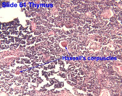
Hassall's corpuscles
| Hassall's corpuscles | |
|---|---|
 Micrograph of a thymic corpuscle. H&E stain. | |
| Details | |
| Part of | Medulla of thymus |
| Anatomical terminology | |
Hassall's corpuscles (also known as thymic bodies) are structures found in the medulla of the human thymus, formed from eosinophilic type VI thymic epithelial cells arranged concentrically. These concentric corpuscles are composed of a central mass, consisting of one or more granular cells, and of a capsule formed of epithelioid cells. They vary in size with diameters from 20 to more than 100μm, and tend to grow larger with age.[1] They can be spherical or ovoid and their epithelial cells contain keratohyalin and bundles of cytoplasmic fibres.[2] Later studies indicate that Hassall's corpuscles differentiate from medullary thymic epithelial cells after they lose autoimmune regulator (AIRE) expression.[3] This makes them an example of Thymic mimetic cells.[4] They are named for Arthur Hill Hassall, who discovered them in 1846.[5][6]
The function of Hassall's corpuscles is currently unclear, and the absence of this structure in the thymus of most murine species (except for the New Zealand White Mouse strain) has previously restricted mechanistic dissection. It is known that Hassall's corpuscles are a potent source of the cytokine TSLP. In vitro, TSLP directs the maturation of dendritic cells, and increases the ability of dendritic cells to convert naive thymocytes to a Foxp3+ regulatory T cell lineage.[7][8] It is unknown if this is the physiological function of Hassall's corpuscles in vivo.
In the past decade, researchers found tissue-specific self-antigens in Hassall's corpuscles and revealed their role in the pathogenesis of diseases such as type 1 diabetes, rheumatoid arthritis, multiple sclerosis, autoimmune thyroiditis, Goodpasture's syndrome, and others. They also discovered that Hassall's corpuscles synthesize chemokines affecting different cell populations in thymic medulla. Despite this, the information on the relationship between Hassall's corpuscles with other cell types of thymic medulla (dendritic, myoid, neuroendocrine cells, thymocytes, macrophages, eosinophils, etc.) remains insufficient and often contradictory. Mechanisms of these relationships and their functional significance are still unclear. The lack of such data does not allow a systemic view of the differentiation processes in the thymus.
References
[edit]![]() This article incorporates text in the public domain from page 1274 of the 20th edition of Gray's Anatomy (1918)
This article incorporates text in the public domain from page 1274 of the 20th edition of Gray's Anatomy (1918)
- ^ Geneser, Finn (1999). Histologi. Munksgaard Danmark. ISBN 87-628-0137-6.
- ^ Dorland's (2012). Dorland's Illustrated Medical Dictionary (32nd ed.). Elsevier. p. 419. ISBN 978-1-4160-6257-8.
- ^ Wang, Xiaoping; et al. (2012). "Post-Aire maturation of thymic medullary epithelial cells involves selective expression of keratinocyte-specific autoantigens". Front. Immunol. 3 (March): 19. doi:10.3389/fimmu.2012.00019. PMC 3310317. PMID 22448160.
- ^ Michelson, Daniel A.; Mathis, Diane (October 2022). "Thymic mimetic cells: tolerogenic masqueraders". Trends in Immunology. 43 (10): 782–791. doi:10.1016/j.it.2022.07.010. PMC 9509455. PMID 36008259.
- ^ Louis Kater: A Note on Hassall's Corpuscles. Contemporary Topics in Immunobiology Volume 2, 1973, pp. 101-109.
- ^ Hassall, A. H., 1846. Microscopic Anatomy of the Human Body in Health and Disease, Highly, London.
- ^ * Watanabe N, Wang Y, Lee H, Ito T, Wang Y, Cao W, Liu Y (2005). "Hassall's corpuscles instruct dendritic cells to induce CD4+CD25+ regulatory T cells in human thymus". Nature. 436 (7054): 1181–5. Bibcode:2005Natur.436.1181W. doi:10.1038/nature03886. PMID 16121185. S2CID 4387582.
- ^ "Old mystery solved, revealing origin of regulatory T cells that 'police' and protect the body"
External links
[edit]- synd/1912 at Whonamedit?
- UIUC Histology Subject 1247
- Histology at KUMC lymphoid-lymph03 "Hassall's Corpuscles"
- Histology image: 61_05 at the University of Oklahoma Health Sciences Center - "Thymus"
- Histology image: 07407loa – Histology Learning System at Boston University - "Lymphoid Tissues and Organs: thymus, Hassall's corpuscles"